Radiologic analyses of the onset of osteogenesis in the grey breasted guinea fowl (Numida meleagris galeata)
Keywords:
Guinea fowl, Long bones, ossification, skull, vertebraeAbstract
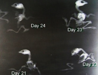
The onset and progress of avian osteogenesis vary with species, and is a tool for teratological and developmental engineering. This work documented the onset and progress of osteogenesis in the grey-breasted guinea fowl based on radiological observations. One hundred and fifty freshly fertilized guinea fowl eggs were incubated using an incubator (Bionics scientific Technologies). Starting from day 7 of incubation, 5 eggs were then cracked daily for the study. Their embryos were harvested routinely and X-rayed. Chronological appearance of primary loci of ossification in the axial and appendicular skeleton were observed. The ossification loci of axial and pectoral limb were presented cranio-caudally, and dorso-ventrally for the pelvic limb. No ossification was observed in the first 14 days of incubation. Skull ossification was first seen on day 16, starting with the bones of the beak. Ossification of the vertebral bones was first observed on day 21 for the cervical vertebrae. The ossification of the vertebral ribs and the sternum were also observed on day 21. Radiograph was unable to reveal the coccygeal and pygostyle throughout the incubation period. The first radiographic evidence of ossification in the appendicular skeleton was that of the long bones of the wings and legs, which appeared by day 15 of incubation. The ossification of the coracoids and clavicle was visible by day 17 of incubation, while that of the pelvic girdle was visible by days 20-23. These results and others were compared with those of other birds and some inferences were derived.

Downloads
Published
How to Cite
Issue
Section
License
Copyright (c) 2023 Sulaiman Olawoye Salami, Chikera Samuel Ibe, Kenechukwu Tobechukwu Onwuama, Zubair Alhaji Jaji, Esther Solomon Kigir

This work is licensed under a Creative Commons Attribution 4.0 International License.


