Computed tomographic (CT) scan study of the anatomy of grasscutter (Thryonomys swinderianus, Temminck 1827): Preliminary observations
DOI:
https://doi.org/10.51607/22331360.2025.74.3.237Keywords:
Anatomy, computed tomography, grasscutter, X-rayAbstract
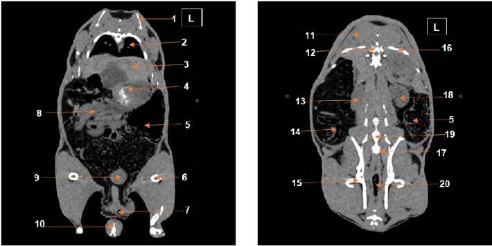
The grasscutter (Thryonomys swinderianus) is an African rodent whose breeding has developed in part due to growing scientific interest. This work is to investigate the internal anatomy of grasscutter (Thryonomys swinderianus) using computed tomography (CT scan). Six grasscutters with an average age of 345 ± 45.63 days and varying sexes and weights, underwent general anesthesia with a ketamine, xylazine mixture and were then subjected to CT scan examination. Volumetric acquisitions were obtained and reconstructed using specific filters: “thoracic or pulmonary tissue,” “abdominal tissue,” and “bone tissue.” Within the thoracic cavity, structures such as the trachea, bronchi, lungs, esophagus, heart, and aorta were identified. In the abdominal cavity, the liver was observed extending transversely from left to right. The stomach, which contained mineral content, as well as the spleen, were clearly identified. The kidneys were also visible, with the left kidney located more cranially than the right. The urinary bladder was identifiable depending on its degree of filling. These same organs have previously been described in rabbits, lemurs, and guinea pigs through CT imaging, supporting comparative anatomical analyses. However, the ureters could not be visualized on the current images. This study represents the first CT scan–based anatomical investigation of the grasscutter and offers valuable insights for applied research in health of this species.
Downloads
Published
How to Cite
Issue
Section
License
Copyright (c) 2025 Aklesso Ataba; Claude Guintard, Mazamaesso Tchaou (Author)

This work is licensed under a Creative Commons Attribution 4.0 International License.