An assessment of cardiac histopathological changes in doxorubicin dose-dependent animal models
DOI:
https://doi.org/10.51607/22331360.2025.74.S1.63Keywords:
Cardiotoxicity, doxorubicin, myocyte injuryAbstract
One effective anthracycline human chemotherapy drug that
is frequently used to treat solid and haematological cancers is
doxorubicin (DOX). The dose-dependent cardiotoxicity of some
medications can result in irreversible heart failure, limiting their
clinical utility. Understanding the pathophysiology and early
detection of DOX-induced cardiac injury is made possible by
animal models, especially rats, using acute models of DOX
cardiotoxicity due to less time-consuming operations. The aim
of this research is to determine a potential cardiotoxic DOX dose
in gender-specific Wistar wild-type rats using light microscopy
for evaluating morphological changes of the heart.
Adult Wistar rats (n=10), including males (n=5) and females
(n=5), were treated with doxorubicin intraperitoneal injection in
different doses (25 mg/kg, 30 mg/kg and 40 mg/kg) per male rat
and female rat, respectively. Rats were sacrificed after 48 hours
and 72 hours for the models of 25 mg/kg and 30 mg/kg, while
the rats of the 40 mg/kg model group were sacrificed 24 hours
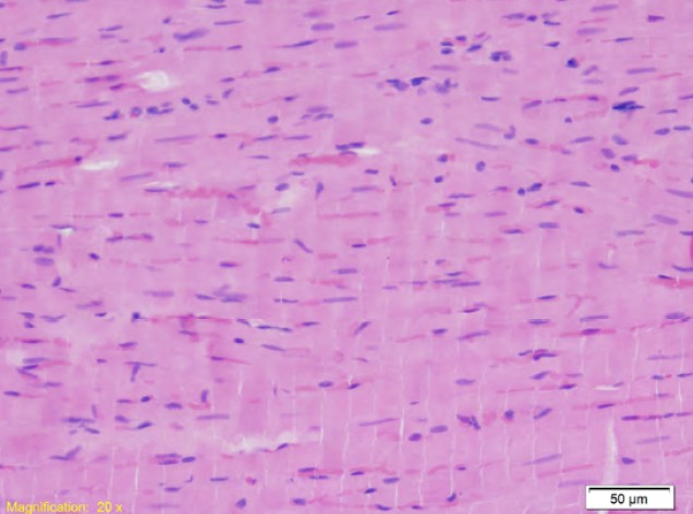
after. The myocardium of the left ventricle is analysed using a
light microscope under magnifications of ten and twenty times.
Male Wistar rats developed more pronounced morphological
changes of the left ventricle compared to female Wistar rats,
resulting in myocardial interstitial oedema and disorganisation
of myocyte architecture.
Male Wistar wild-type rats develop a more aggressive form of
acute cardiotoxicity caused by doxorubicin compared to female
Wistar wild-type rats.
Downloads
Published
How to Cite
Issue
Section
License
Copyright (c) 2025 Rijad Jahić, Edina Lazović Salčin, Muhamed Katica, Almir Fajkić

This work is licensed under a Creative Commons Attribution 4.0 International License.