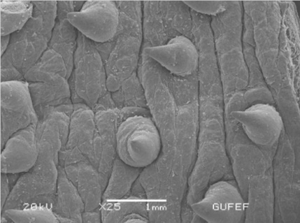
Stereomicroscopic and scanning electron microscopic observations of the forestomach mucosa of Akkaraman sheep fed with straw-concentrate diet
DOI:
https://doi.org/10.51607/22331360.2023.72.3.302Keywords:
Intermediate type eaters, sheep, forestomach components, SEMAbstract
This research was conducted to observe the mucosal surface of the forestomach components of seven Akkaraman sheep fed with straw-concentrate diet using stereomicroscopy and SEM. The samples were obtained from the animals fed 60% straw and 40% concentrate, and slaughtered in a local slaughterhouse. The results revealed that the ruminal papillae were present on all of the surface of the rumen except the ruminal pila. Their shape varied considerably in shape and size from a short lingual to a long and wide leaf-like forms. Some papilla showed asymmetrical doubled-apices. The cells in the ruminal mucosa were mostly intermediate-type cells. Occasionally, their shapes resembled balloon-type cells. Reticular cristas in the reticulum and omasal lamina in the omasum were also displayed clearly, but certain papilla types and their peculiarities were not eminent at macroscopical level. At microscopical level, there were papilla- type structures present on the omasal lamina. They had smooth surface, mostly possessing one, occasionally double ridges. The epithelial scrap layer formed by the horny cells was eminent, indicating regeneration of the papilla. The results have shown the characteristics of the animals fed with strawconcentrate diet.
References
Downloads
Published
Issue
Section
License
Copyright (c) 2023 Şükrü Hakan Atalgin, İbrahim Kürtül, Lütfi Takçi

This work is licensed under a Creative Commons Attribution 4.0 International License.