Gross anatomy and histology of the urinary system of the spur winged goose (Plectropterus gambensis)
DOI:
https://doi.org/10.51607/22331360.2024.73.1.53Keywords:
Avian, cloaca, kidney, ureter, waterfowlAbstract
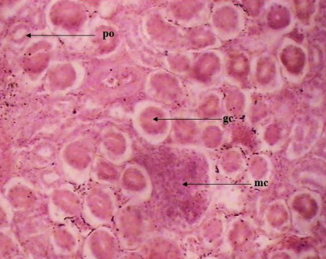
Damaturu, Yobe State, Nigeria and transported to the Gross and Histology Postgraduate Research Laboratory of the Department of Veterinary Anatomy, University of Maiduguri, Nigeria. The birds were acclimatized for 10 days, before chemically euthanized. The abdominal cavity of the birds was exteriorized to grossly observe the kidneys, ureters and cloaca before harvested for microscopic study. Grossly, the kidney appeared brownish, dorso-laterally flattened with three distinct lobes. The ureter appears as a tubular vessel emanating from the kidneys and enters the cloaca at the dorsomedial aspect. The cloaca was observed as a common organ that connects the urinary, reproductive and digestive system. Histologically, the kidney parenchyma was divided into renal cortex and medulla, containing the central vein and renal corpuscles respectively. The lamina epithelia of the ureter were lined with a pseudostratified columnar epithelium containing loose connective tissues and lymphoid cells. The mucosa of the cloaca possesses a short and slender villi projection. Currently, there is no baseline information regarding the anatomic characteristics of the urinary system of the spur winged goose. Therefore, this present study serves as firsthand information on the anatomy of the urinary system of the spur winged geese.
Downloads
Published
How to Cite
Issue
Section
License
Copyright (c) 2024 Yagana Bukar Majama, Ali Musa Wulgo, Mohammed Kachalla Malah, Innocent David Kwabe

This work is licensed under a Creative Commons Attribution 4.0 International License.